Santosh K. Padala1 MD, José-Angel Cabrera2 MD, Kenneth A. Ellenbogen1 MD
1 Division of Cardiology, Pauley Heart Center, Virginia Commonwealth University, VA, USA
2 Unidad de Arritmias, Departamento de Cardiología, Hospital Universitario Quirónsalud Salud Madrid.
Abstract
The specialized cardiomyocytes that constitute the conduction system in the human heart initiate the
electric impulse and result in rhythmic and synchronized contraction of the atria and ventricles.
Although the atrioventricular (AV) conduction axis was described more than a century ago by Sunao
Tawara, the anatomic pathway for propagation of impulse from atria to the ventricles has been a topic
of debate for years. Over the past 2 decades there has been a resurgence of conduction system pacing
(CSP) by implanting pacing leads in the His bundle region in lieu of chronic right ventricular pacing
that is associated with worse clinical outcomes. The inherent limitations of implanting the leads in the
His bundle region has led to the emergence of left bundle branch area pacing in the past 3 years as an
alternative strategy for CSP. The clinical experience from performing CSP has helped
electrophysiologists gain deeper insight into the anatomy and physiology of cardiac conduction
system. This review details the anatomy of the cardiac conduction system and highlights some of the
recently published articles that aid in better understanding of the AV conduction axis and its
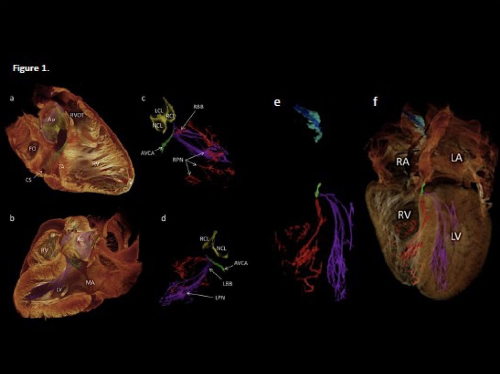
variations, the knowledge of which is critical for CSP. The remarkable evolution in technology has
led to visualization of the cardiac conduction system using non-invasive, non-destructive highresolution
contrast enhanced micro-computed tomography imaging that may aid in future CSP. We
also discuss from anatomical perspective the differences seen clinically with His bundle pacing and
left bundle branch area pacing.